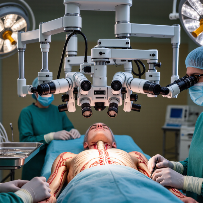
| What is a spine surgery microscope? | An operating microscope designed to provide magnified, illuminated views of the surgical field. | It can help the surgeon see small anatomical structures, including nerve roots and fine tissue planes, during selected procedures. | It supports visualization but does not replace surgical training, clinical judgment, or other imaging tools. |
| Magnification | Optical magnification makes the operative area appear larger to the surgeon. | A closer view can assist with precise work in a confined surgical corridor, such as during some decompression or microdiscectomy procedures. | The useful level of magnification depends on the task, field of view, and surgeon preference. |
| Illumination | Integrated lighting directs light into the operative field. | Focused illumination can improve visibility in deeper areas that may be difficult to light directly. | Lighting quality and positioning should be appropriate for the procedure and working distance. |
| Depth perception | Binocular optics provide a stereoscopic view when the microscope is properly set up. | A three-dimensional view can help the surgeon judge spatial relationships while working around delicate structures. | The benefit depends on correct adjustment, a clear line of sight, and the operator’s experience. |
| Typical applications | Microscope use may be considered for procedures involving fine dissection or a limited operative field. | Examples can include selected lumbar microdiscectomies, decompressions, and operations involving spinal tumors or intradural structures. | Use varies by procedure, surgeon, patient factors, and local practice; it is not required for every spine operation. |
| Ergonomics and positioning | The microscope can be positioned to provide a stable view while the surgeon works through the operative approach. | Appropriate positioning may help the surgeon maintain a comfortable viewing posture during fine work. | Setup and repositioning take time, and the microscope must not obstruct access or other equipment. |
| What it does not do | A microscope improves the surgeon’s optical view of the field; it does not independently identify anatomy or guide instruments. | Fluoroscopy, navigation, or other imaging may still be used when clinically indicated, particularly for procedures requiring instrument or implant localization. | The appropriate combination of visualization and imaging depends on the operation and the patient’s needs. |
| Choosing a microscope | Relevant factors include optical clarity, illumination, working distance, maneuverability, and compatibility with the operating room. | A suitable setup should support the procedures performed and allow the surgical team to work safely and efficiently. | Selection should account for the surgeon’s workflow, staff training, maintenance, and facility requirements. |